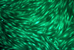
Autophagy is a lysosomal degradation pathway for cytoplasmic material and is activated during cellular stress. Upon induction by a stress signal such as viral infection or amino acid starvation, the first step in autophagy is the formation of an autophagosome. The most published autophagosome marker protein is MAP LC3, which associates to the inner and outer limiting membranes of the autophagosome.
LC3 is a small 16-18 kDa protein that is found in 3 forms within cells. The LC3 pro-form of 125 amino acids becomes cleaved to a 120 amino acid form known as LC3-I. Upon binding to phosphatidylethanolamine (PE), the LC3-II form is generated. LC3-I is found mainly in the cytosol, and LC3-II is found mainly in the membrane fraction. Both LC3-I and LC3-II are seen in healthy cells, but upon induction of autophagy the LC3-II proportion increases as it associates with the membranes of the autophagosomes.
Our Autophagy ELISA Kit (LC3-II Quantitation) uses a selective permeabilization procedure to remove the cytosolic pro-LC3 and LC3-I and retain the autophagosome membrane-bound LC3-II. The quantity of LC3-II in unknown lysates is determined by comparison to a known LC3 standard curve.
Awad, A.M. et al. (2023). Ameliorative effect of montelukast against STZ induced diabetic nephropathy: targeting HMGB1, TLR4, NF-κB, NLRP3 inflammasome, and autophagy pathways. Inflammopharmacology. doi: 10.1007/s10787-023-01301-1.