Cell Adhesion Assays, ECM Array

Cell Adhesion Assays, ECM Array
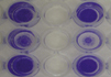
  • Full quantitation of cell adhesion with no manual cell counting
  • Fluorometric or colorimetric detection
  • Plates precoated with a uniform substrate layer of a single ECM protein in each row: Collagen I, Collagen IV, Fibrinogen, Fibronectin, and Laminin


Frequently Asked Questions about this product

Email To BuyerPrint this PageCopy Link
Ordering

Please contact your distributor for pricing.

CytoSelect™ 48-Well Cell Adhesion Assay, ECM Array
Catalog Number
CBA-070
Size
48 assays
Detection
Colorimetric
Manual/Data Sheet Download
SDS Download
Price
$515.00
CytoSelect™ 48-Well Cell Adhesion Assay, ECM Array
Catalog Number
CBA-071
Size
48 assays
Detection
Fluorometric
Manual/Data Sheet Download
SDS Download
Price
$555.00
CytoSelect™ 48-Well Cell Adhesion Assay, ECM Array
Catalog Number
CBA-070-5
Size
5 x 48 assays
Detection
Colorimetric
Manual/Data Sheet Download
SDS Download
Price
$2,275.00
CytoSelect™ 48-Well Cell Adhesion Assay, ECM Array
Catalog Number
CBA-071-5
Size
5 x 48 assays
Detection
Fluorometric
Manual/Data Sheet Download
SDS Download
Price
$2,430.00
Product Details

Cell adhesion is a complex process involved in migration/invasion, embryogenesis, wound healing and tissue remodeling. Cells adhere to the extracellular matrix, forming complexes with cytoskeleton components that can affect cell motility, differentiation, proliferation, and survival.

Our CytoSelect™ 48-Well Cell Adhesion Assays provide a fully quantitative method for the evaluation of cell adhesion. The 48-well plate is precoated with your choice of single ECM protein in each of the first 5 rows, with the last row provided as a negative control. Just follow a simple protocol:

  1. Cells are seeded onto the substrate.
  2. Adherent cells attach, while non-adherent cells are washed away.
  3. Adherent cells are quantified on a standard plate reader or fluorometer.

CytoSelect™ 48-Well Cell Adhesion Assay, ECM Array. Serum starved cells from three different cell lines were allowed to attach to the ECM-coated 48-well plate for 1 hr at 100,000 cells/well. Adherent cells were stained according to the assay protocol. BSA = Bovine Serum Albumin. VN = Vitronectin. LN = Laminin. FN = Fibronectin. Col = Collagen.

Recent Product Citations
  1. Liu, M.X, et al. 2023. MiR-410-3p suppresses primary gastric cancer and exosomes regulate endogenous expression of miR-410-3p. Am J Cancer Res. 13(6):2670-2680 (#CBA-070).
  2. García-García, T. et al. (2022). Impairment of antiviral immune response and disruption of cellular functions by SARS-CoV-2 ORF7a and ORF7b. iScience. doi: 10.1016/j.isci.2022.105444 (#CBA-070).
  3. Khan, A.A. et al. (2022). Determination of anticancer potential of a novel pharmacologically active thiosemicarbazone derivative against colorectal cancer cell lines. Saudi Pharm J. doi: 10.1016/j.jsps.2022.03.011 (#CBA-070).
  4. Lepsenyi, M. et al. (2021). CXCL2-CXCR2 axis mediates αV integrin-dependent peritoneal metastasis of colon cancer cells. Clin Exp Metastasis. doi: 10.1007/s10585-021-10103-0 (#CBA-071).
  5. Park, S. et al. (2021). Production of a Foot-and-Mouth Disease Vaccine Antigen Using Suspension-Adapted BHK-21 Cells in a Bioreactor. Vaccines (Basel). 9(5):505. doi: 10.3390/vaccines9050505 (#CBA-070).
  6. Takeshita, Y. et al. (2021). Angiopoietin-like protein 2 decreases peritoneal metastasis of ovarian cancer cells by suppressing anoikis resistance. Biochem Biophys Res Commun. 561:26-32. doi: 10.1016/j.bbrc.2021.05.008 (#CBA-070).
  7. Potter, J.A. et al. (2021). Collagen I Modifies Connexin-43 Hemichannel Activity via Integrin α2β1 Binding in TGFβ1-Evoked Renal Tubular Epithelial Cells. Int J Mol Sci. 22(7):3644. doi: 10.3390/ijms22073644 (#CBA-070).
  8. Liu, S.Y. et al. (2021). Evaluation of Nanotargeted 111In-Cyclic RGDfK-Liposome in a Human Melanoma Xenotransplantation Model. Int J Mol Sci. 22(3):1099. doi: 10.3390/ijms22031099 (#CBA-070).
  9. Iosef, C. et al. (2020). Quantitative proteomics reveal lineage-specific protein profiles in iPSC-derived Marfan syndrome smooth muscle cells. Sci Rep. 10(1):20392. doi: 10.1038/s41598-020-77274-w (#CBA-070).
  10. Karatsai, O. et al. (2020). Combinatory Treatment of Canavanine and Arginine Deprivation Efficiently Targets Human Glioblastoma Cells via Pleiotropic Mechanisms. Cells. 9(10):E2217. doi: 10.3390/cells9102217 (#CBA-070).
  11. Celentano, A. et al. (2020). Kava constituents exert selective anticancer effects in oral squamous cell carcinoma cells in vitro. Sci Rep. 10(1):15904. doi: 10.1038/s41598-020-73058-4 (#CBA-070).
  12. Jha, S.K. et al. (2020). Enhanced antitumor efficacy of bile acid-lipid complex-anchored docetaxel nanoemulsion via oral metronomic scheduling. J Control Release. 328:368-394. doi: 10.1016/j.jconrel.2020.08.067 (#CBA-070).
  13. Kanda, M. et al. (2020). Therapeutic monoclonal antibody targeting of neuronal pentraxin receptor to control metastasis in gastric cancer. Mol Cancer. 19(1):131. doi: 10.1186/s12943-020-01251-0 (#CBA-071).
  14. Bagdonaite, I. et al. (2020). O-glycan initiation directs distinct biological pathways and controls epithelial differentiation. EMBO Rep. doi: 10.15252/embr.201948885 (#CBA-070).
  15. Okada, J. et al. (2020). Dapagliflozin Inhibits Cell Adhesion to Collagen I and IV and Increases Ectodomain Proteolytic Cleavage of DDR1 by Increasing ADAM10 Activity. Molecules. 25(3). pii: E495. doi: 10.3390/molecules25030495 (#CBA-070).
  16. Nakamura, S. et al. (2020). KCNJ15 Expression and Malignant Behavior of Esophageal Squamous Cell Carcinoma. Ann Surg Oncol. doi: 10.1245/s10434-019-08189-8 (#CBA-071).
  17. Liu, L.Q. et al. (2019). MiR-92a antagonized the facilitation effect of extracellular matrix protein 1 in GC metastasis through targeting its 3'UTR region. Food Chem Toxicol. 133:110779. doi: 10.1016/j.fct.2019.110779 (#CBA-070).
  18. Hou, S. et al. (2019). Integrin α5 promotes migration and cisplatin resistance in esophageal squamous cell carcinoma cells. Am J Cancer Res. 9(12):2774-2788 (#CBA-070).
  19. Uno, Y. et al. (2019). Expression, Function, and Prognostic Value of MAGE-D4 Protein in Esophageal Squamous Cell Carcinoma. Anticancer Res. 39(11):6015-6023. doi: 10.21873/anticanres.13807 (#CBA-070).
  20. Umeda, S. et al. (2019). Fraser extracellular matrix complex subunit 1 promotes liver metastasis of gastric cancer. Int J Cancer. doi: 10.1002/ijc.32705 (#CBA-070).
  21. Hirano, K. et al. (2019). LacdiNAcylation of N-glycans in MDA-MB-231 human breast cancer cells results in changes in morphological appearance and adhesive properties of the cells. Histochem Cell Biol. doi: 10.1007/s00418-019-01822-3 (#CBA-070).
  22. Ding, L. et al. (2019). Perturbed myoepithelial cell differentiation in BRCA mutation carriers and in ductal carcinoma in situ. Nat Commun. 10(1):4182. doi: 10.1038/s41467-019-12125-5 (#CBA-070).
  23. Birtley, J.R. et al. (2019). Inactivating mutations and X-ray crystal structure of the tumor suppressor OPCML reveal cancer-associated functions. Nat Commun. 10(1):3134. doi: 10.1038/s41467-019-10966-8 (#CBA-070).
  24. Sohn, H.J. et al. (2019). Cellular characterization of actin gene concerned with contact-dependent mechanisms in Naegleria fowleri. Parasite Immunol. e12631. doi: 10.1111/pim.12631 (#CBA-070).
  25. Babchia, N. et al. (2019). The bidirectional crosstalk between metastatic uveal melanoma cells and hepatic stellate cells engenders an inflammatory microenvironment. Exp Eye Res. 181:213-222. doi: 10.1016/j.exer.2019.02.012 (#CBA-070).
  26. Sawaki, K. et al. (2019). Level of Melanotransferrin in Tissue and Sera Serves as a Prognostic Marker of Gastric Cancer. Anticancer Res. 39(11):6125-6133. doi: 10.21873/anticanres.13820 (#CBA-071).
  27. Maxwell, J.T. et al. (2019). Electrical stimulation of pediatric cardiac-derived c-kit+ progenitor cells improves retention and cardiac function in right ventricular heart failure. Stem Cells. doi: 10.1002/stem.3088 (#CBA-071).
  28. Uno, Y. et al. (2019). Increased Expression of DNAJC12 is Associated with Aggressive Phenotype of Gastric Cancer. Ann Surg Oncol. 26(3):836-844. doi: 10.1245/s10434-018-07149-y (#CBA-071).
  29. Zohra, F. T. et al. (2015). Functional behavior and gene expression of magnetic nanoparticle-loaded primary endothelial cells for targeting vascular stents. Nanomedicine (Lond). 10:1391-1406 (#CBA-071).
  30. Kim, S.W. et al. (2013). Cardiac Stem Cells with Electrical Stimulation Improve Ischaemic Heart Function through Regulation of Connective Tissue Growth Factor and miR-378. Cardiovasc Res. 10.1093/cvr/cvt192 (#CBA-071).